

Drug discovery has always had a translation problem. A compound can look very good in a cell-based assay, perform well enough in animals, and still fail once it reaches clinical testing. Sometimes the issue is efficacy. Sometimes it is toxicity or exposure. Often, human biology is simply more complex than the model used earlier in development.
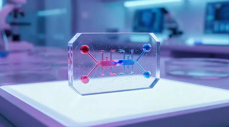
This is where an organ-on-a-chip platform may help. These human-cell-based microphysiological systems are part of new approach methodologies (NAMs), alongside 3D cell models and organoids. An organ-on-a-chip platform places living cells inside a small engineered device where fluid flow, tissue organisation, cell-cell interactions, and mechanical conditions can be controlled. The aim is not to build a tiny, complete human organ. It is to reproduce enough of the organ’s behaviour to answer a useful drug-development question.
(AI-generated image for illustration)
Why microphysiological systems go beyond 2D cell culture
Conventional 2D culture is still extremely useful. It is inexpensive, relatively easy to reproduce, and suitable for screening large numbers of compounds. The problem is that cells growing as a flat monolayer behave differently from cells living inside a tissue.
In the body, cells interact with neighbouring cells and the extracellular matrix. They experience gradients in oxygen, nutrients, signalling molecules, and drug concentrations. Tissue structure also determines how a compound moves from one compartment to another. These features are mostly absent in standard monolayer culture. The webinar highlighted that this can lead to very different exposure conditions and biological responses compared with a more tissue-like system.
Three-dimensional models partly address this. Spheroids allow cells to organise in space. Organoids can recreate some tissue-specific structures and functions. Organ-on-chip systems add another dimension because they can introduce flow, different tissue compartments, and sometimes mechanical forces. In practice, the models are becoming more sophisticated step by step, from simple 3D cultures to microphysiological systems and, eventually, linked multi-organ platforms.
More complexity is not always better, though. If a simple assay answers the question, there is little reason to use a chip. These platforms become more useful when barrier properties, flow, tissue architecture, repeated exposure, or interaction between different cell types are likely to affect the result.
Where an organ-on-a-chip platform fits into DMPK
DMPK is one area where this becomes particularly relevant.
Absorption, distribution, metabolism, and excretion are not isolated chemical processes. They depend heavily on tissue physiology. A drug must cross barriers, encounter transporters and enzymes, distribute between compartments, and eventually be metabolised or cleared.
A gut-on-chip, for example, can be used to investigate intestinal transport. Liver systems can help examine drug metabolism, metabolite formation, and liver toxicity. Kidney-on-chip models can be useful for studying tubular transport and renal handling. Blood-brain barrier models are being explored to understand whether compounds can enter the central nervous system. Multi-organ models go one step further by asking how processes in one tissue influence what happens in another. These are among the applications discussed in the webinar in relation to drug discovery and DMPK.
It would be premature to see these systems as replacements for conventional ADME or in vivo studies. Their value is more likely to be in filling specific gaps. If a conventional assay says a compound has poor permeability, an organ-on-chip model may help explain why. If toxicity appears only after repeated exposure, a continuously perfused model may provide useful mechanistic information that a short static assay cannot.
Skin-on-chip as a practical organ-on-chip model
Skin is a particularly interesting case because its function depends so strongly on its physical organisation.
The epidermis forms the main barrier. Beneath it lies the dermis, with fibroblasts and extracellular matrix. Drug transport through skin depends on how well these layers are formed, how tightly the cells are connected, and how the compound interacts with the barrier.
Commercial reconstructed epidermal and full-thickness skin models are already used in research. But the webinar points out several limitations for some applications, including limited physiological nutrient transport, the absence of vascular-like function, and constraints in systemic toxicity and time-dependent permeability studies.
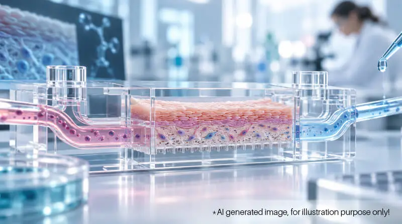
The organ-on-a-chip platform presented in the webinar used a microfluidic design containing an upper cell-culture chamber, a porous membrane, and a lower channel through which culture medium could flow. Human dermal fibroblasts and keratinocytes were cultured together in three dimensions under continuous perfusion.
But putting two types of skin cells inside a device does not automatically make it a skin model. The more important question is whether those cells organise and mature in a skin-like way.
That was assessed using microscopy, histology, and tissue-specific markers. Distinct epidermis-like and dermis-like regions were observed. Fibronectin and vimentin supported the dermal fibroblast phenotype. E-cadherin indicated cell-cell junction formation in the epidermal compartment. Cytokeratin 14 marked basal keratinocytes, while cytokeratins 1 and 10, involucrin, and filaggrin provided evidence of keratinocyte differentiation and progressive maturation of the epidermal layer.
This is important because a permeability model is only useful if the barrier itself has formed properly.
Does the barrier actually work?
The group measured barrier function using transepithelial electrical resistance, or TEER. In simple terms, TEER tells us how difficult it is for electrical current to pass across a cellular barrier. A higher value usually indicates tighter junctions and better barrier integrity.
The 3D skin co-culture showed higher TEER than simpler epidermal or dermal cultures. The reported TEER value was around 1,926 Ω·cm², within the range cited for established in vitro skin models. FITC-dextran permeability measurements also suggested that the barrier remained relatively stable over time.
The model was then tested with caffeine, salicylic acid, hydrocortisone, and clotrimazole. These compounds differ in their physicochemical behaviour and produced different permeability profiles. That is exactly what is needed from a useful model. If every compound behaves in the same way, the system tells us very little.
The device itself can become part of the problem
One point from the webinar that deserves more attention is the material used to build the chip.
Early devices used PDMS, a material commonly used in microfluidics. It is convenient and well established, but it is not perfect. PDMS can allow evaporation, swell in contact with some solvents, show variability between batches, and adsorb certain drug molecules. That last issue matters in DMPK studies because the concentration measured in the experiment may no longer reflect the amount originally added.
The group therefore explored a selective laser sintering, or SLS, 3D-printed device made using an ABS-like polymer. The intention was to improve mechanical strength, chemical resistance, reproducibility, and scalability. A six-well format with a common inlet and separate outlets was developed for parallel experiments. They also tested 3D bioprinting of the skin co-culture. In the experiments shown, the bioprinted construct produced better barrier properties than the manually seeded version.
For an organ-on-a-chip platform to be useful beyond a research demonstration, these engineering details matter. Device design, materials, manufacturing, fluidics, and analytical compatibility can determine whether the biological result is trustworthy.
New approach methodologies (NAMs): where does this go next?
Organ-on-chip systems are becoming more relevant as interest in new approach methodologies grows, but there is still a large gap between an interesting academic model and a routinely useful drug-development platform.
Reproducibility across laboratories matters. So does scale-up. The model must remain stable over time, perform consistently between batches, and respond appropriately to reference compounds. The context of use also has to be clear. A model validated for skin permeability cannot automatically be assumed to predict systemic toxicity.
For drug discovery organisations such as Syngene, the real opportunity will probably come from combining these models with existing DMPK, pharmacology, bioanalysis, toxicology, imaging, and computational approaches. Organ-on-chip data alone may not answer the whole question, but they can add a more human-relevant layer to the evidence already being generated.
The skin-on-chip work presented in the webinar shows how that could happen. Human cells, 3D tissue organisation, controlled flow, barrier measurements, drug permeability, material science, and device engineering were all brought into the same experimental system.
The field probably does not need the most complicated chip possible. It needs an organ-on-a-chip platform that is biologically relevant, technically reliable, and useful enough to change a real development decision. That is the harder problem, and also the more important one.